OPG is a panoramic dental X-ray that captures the entire upper and lower jaw in a single image. In Australian dentistry, it usually means orthopantomogram, and it gives a wide view of all teeth, the jaw joints, and nearby structures.
A lot of people see “OPG” on a referral slip and pause for a second. They're not always sure whether it's a scan, a test, or just paperwork, and that confusion is understandable because the acronym has more than one meaning outside dentistry.
What the Letters OPG Stand For in Dentistry
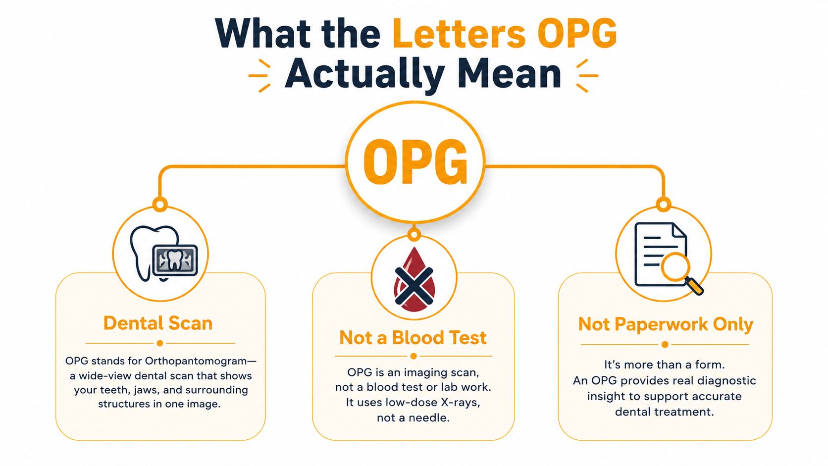
A patient might be standing at reception, holding a referral that says OPG, and wondering if it refers to a test, a form, or something else entirely. In a dental clinic, the meaning is much simpler. OPG stands for orthopantomogram, which is a panoramic dental X-ray that shows the upper jaw, lower jaw, all teeth, the TMJ region, and surrounding structures in one broad image. Darlinghurst Dental notes that it is less detailed than an intra-oral X-ray, but very useful for spotting broader changes such as caries, periodontal bone loss, abscesses, cysts, and other jaw pathology, and for planning extractions and orthodontic treatment.
Why the acronym causes confusion
Outside dentistry, OPG can also mean Osteoprotegerin, Ontario Power Generation, or the Office of the Public Guardian, so it is easy to land on the wrong meaning if you have come from a hospital letter, a public-sector document, or a general web search Acronyms and Abbreviations. That is why a clear patient explanation should begin with the dental meaning, then clear away the other possibilities.
Practical rule: if the document mentions teeth, jaws, orthodontics, or extraction planning, OPG almost certainly means the dental X-ray.
For people who like to keep track of appointments and reminders in one place, it can help to browse the mobile loyalty app used by some practices for convenient access to visit information and updates. That kind of tool does not change the scan itself, but it does make the patient journey easier to follow.
In dentistry, OPG means a panoramic X-ray of the jaws and teeth, and that is the meaning this article uses from here on.
How the Panoramic Image Is Built
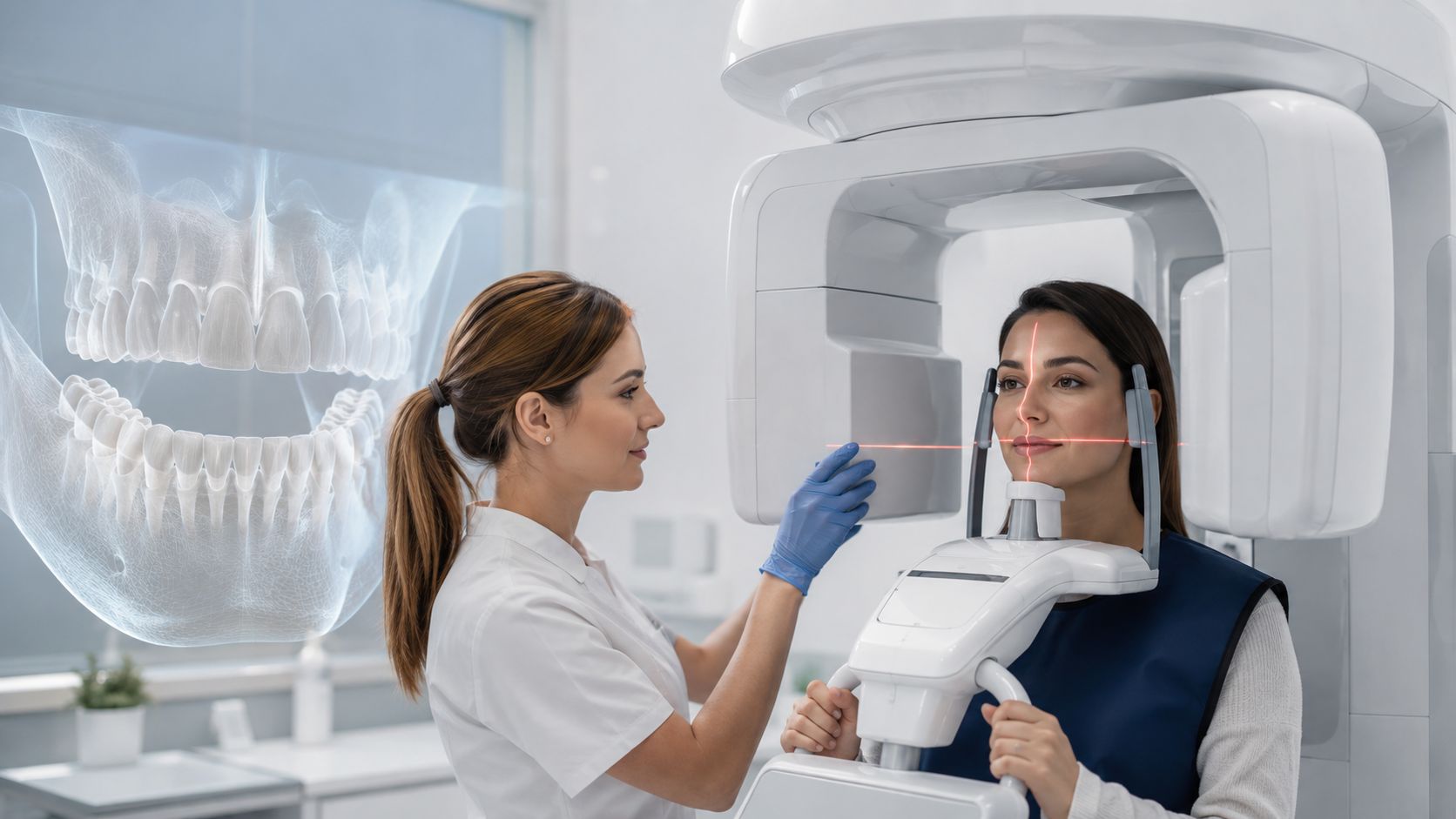
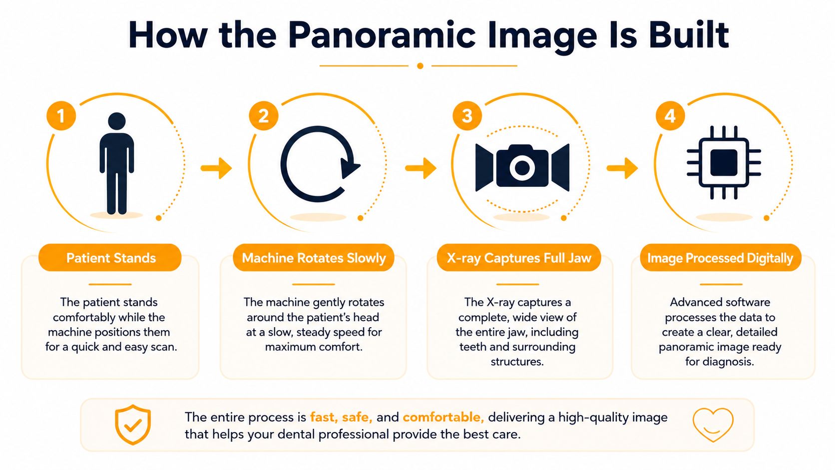
An OPG works a bit like a camera sweeping around your face, except it is designed to map the jaws rather than capture a skin-level snapshot. The machine uses a rotating X-ray source and a moving detector to build a broad image from ear to ear, and the result depends on the jaw sitting inside the correct curved zone, often called the focal trough Radiopaedia.
The focus window idea
A simple way to understand the focal trough is as a focus window. If your teeth and jaws line up with that window, the image comes through clearly. If they sit too far forward, too far back, or slightly tilted, the result can look blurred or stretched because panoramic imaging is a tomographic projection, not a close-up of one tooth.
The positioning step matters more than many patients realise. The team centres you carefully, often using the Frankfort horizontal line parallel to the floor and laser guides, so the jaws stay in the correct plane while the arm moves around you. The operating range is commonly reported at 70–80 kVp and 8–15 mA over a few seconds, which sounds technical, but for the patient it usually feels like standing still while the machine does the work Radiopaedia. Small alignment errors can blur the final image, which is why the radiographer's prompts matter so much.
That trade-off is deliberate. An OPG gives up fine detail so it can show a much bigger picture, and that is what makes it useful for broad screening and treatment planning. If you want the chair-side explanation of a very different close-up tool, an intraoral camera overview shows how dentists use detailed images inside the mouth.
For readers who are comparing scan choices or trying to understand how dentists present local care online, the playbook for ranking in Maps can explain why nearby clinics often show up first in search.
OPG Compared to Other Dental Imaging
OPG sits in the middle of dental imaging. It's broader than a tiny close-up, but it isn't the answer for every diagnostic question. That's why dentists choose it for some jobs and use other scans for different ones.
Where each scan fits
Intraoral X-rays are the sharpest option for one tooth or a small group of teeth. Dentists use them when they need to inspect decay between teeth, root tips, or a localised problem in fine detail. Cephalometric views are side-profile images, and they're especially useful in orthodontic planning because they show how the jaws line up from the side. Cone beam CT, or CBCT, goes further again because it gives three-dimensional bone information, which is valuable for complex implant and surgical cases.
A practical way to read the choice is this. If the dentist wants the whole picture, OPG is often the right first step. If they need to zoom in on one tooth, intraoral imaging makes more sense. If they need 3D bone measurements, CBCT is the better tool.
| Imaging Type | What It Shows Best | Typical Use Case | Detail Level |
|---|---|---|---|
| OPG | Whole jaws, all teeth, jaw joints, broad anatomy | Wisdom teeth checks, orthodontic planning, implant overview | Broad, less detailed |
| Intraoral X-ray | One tooth or a small area | Decay, root issues, local pain | High detail |
| Cephalometric view | Side-profile jaw relationship | Orthodontic assessment | Moderate |
| CBCT | Three-dimensional bone and anatomy | Implants, surgical planning, complex cases | Very detailed |
A clinic that handles referrals efficiently often needs more than one imaging pathway. That same workflow thinking also matters in local search and patient access, which is why resources like a playbook for ranking in Maps can be relevant for practices organising how patients find services online.
At The Smile Spot, the choice of scan is tied to the clinical question, not to a one-size-fits-all routine. If the question is broader than one tooth but not complex enough for 3D imaging, an OPG usually gives the dentist the clearest starting point.
When Dentists Order an OPG
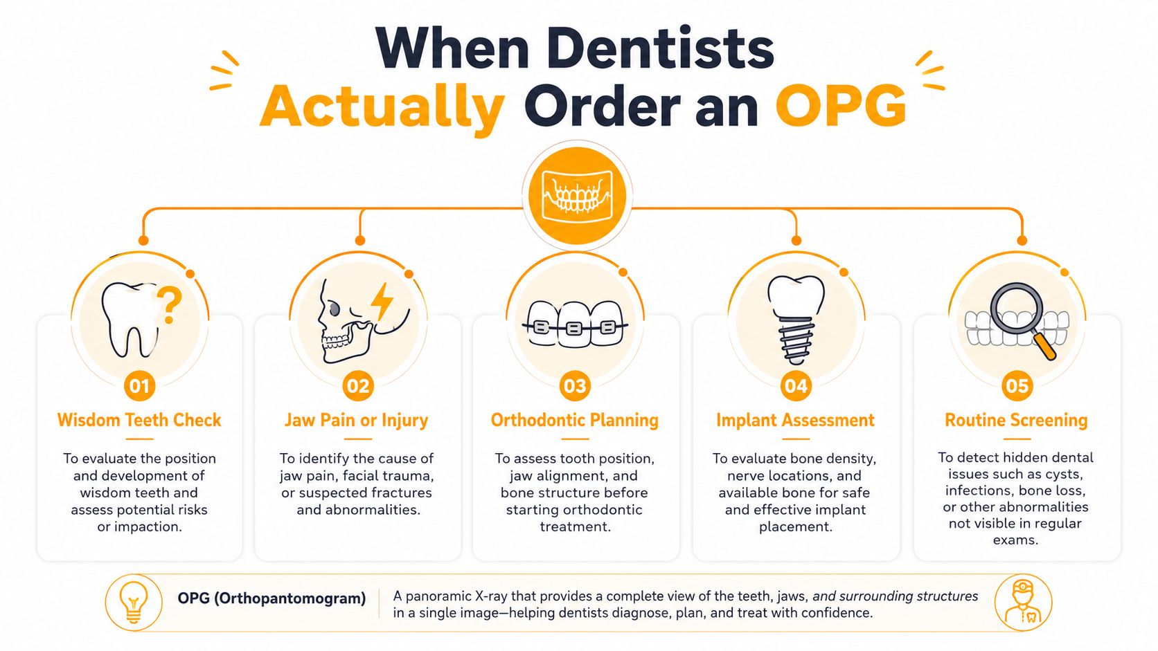
A patient might come in for one sore tooth, yet the dentist needs to understand the whole mouth before choosing the next step. An OPG gives that wider view in a single image, showing the number, position, and eruption status of all teeth, including unerupted teeth, so it often becomes the first scan before treatment begins MCA Dental.
Five common reasons
A teenager being assessed for braces may need an OPG so the orthodontist can check eruption patterns and jaw relationships before treatment starts. An adult with possible wisdom tooth problems may need one to see whether the teeth are impacted, growing in a risky direction, or sitting near other structures. Someone planning a single implant may need a broad look at bone height and the nearby maxillary sinus or nerve pathways before treatment is mapped out. In a more complex extraction, the dentist may want the wider view first so they can judge root position and surrounding anatomy. When there's a concern about cysts, periodontal bone loss, abscesses, or other jaw pathology, the scan can reveal changes across the whole mouth rather than one isolated spot Darlinghurst Dental MCA Dental.
- Wisdom teeth assessment: A young adult with sore back molars might need an OPG before a removal discussion.
- Orthodontic planning: A teenager waiting for braces may need the scan to check eruption and spacing.
- Implant assessment: An adult replacing a missing tooth may need the broader jaw view before implant planning.
- Complex extractions: A dentist may order it when a tooth looks difficult to remove safely.
- Broad screening: A patient with unexplained jaw symptoms may need the image to look for hidden pathology.
If the whole mouth matters more than one tooth, an OPG is often the first image that makes the treatment plan clearer.
For readers who are trying to understand wisdom tooth care in a practical way, a wisdom tooth removal guide sits naturally alongside this topic, because wisdom teeth are one of the most common reasons an OPG gets ordered.
What Happens Before, During, and After the Scan
A lot of patients expect an OPG to feel more complicated than it is. In practice, it is usually a short visit, with the team setting everything up first and the scan itself taking only a moment once you are positioned correctly.
Before the scan
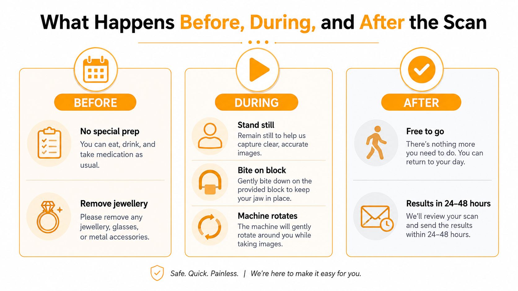
The dentist or radiographer usually starts with a few practical questions. They may ask about pregnancy, any recent imaging, and whether you have jaw, neck, or movement issues that could affect how you stand or sit during the scan. You will also be asked to remove jewellery, glasses, dentures, and hair clips so nothing blocks the image.
That part often sounds more involved than it feels. You stand or sit in the machine, the team guides your head into the right position, and you may bite gently on a small mouthpiece so your teeth stay lined up. Then the arm of the machine moves around your head while you keep still.
During and after the scan
The exposure is brief, and the main job during it is to stay relaxed and still while the machine turns. The image appears on the screen soon after, and the dentist checks it with you or later as part of your treatment planning Radiopaedia.
Patients can usually drive, eat, and go back to normal activities straight away. There is no recovery time, no numbness, and no special aftercare for a standard OPG. In a modern practice, the same imaging room may also support cephalometric and cone beam CT scans, so if a more detailed image is needed later, the process can often stay in the same place Health Facility Guidelines.
For families fitting imaging into routine care, it helps when the scan sits inside an ordinary appointment. A check-up and clean guide can also make the visit feel less unfamiliar before you arrive.
Safety, Radiation Dose, and How Often You Can Have One
The question patients ask most often is simple. Is it safe? For a standard OPG, dentists use a low dose of ionising radiation, and the scan is designed to answer a specific diagnostic question rather than be repeated routinely at every check-up. That's why the decision is based on whether the image will change the treatment plan.
How dentists keep it conservative
The technique uses carefully controlled settings, and the team follows the ALARA principle, which means keeping radiation as low as reasonably achievable. Lead protection may be used where appropriate, and the staff will be especially careful with children, pregnant patients, and people who have had recent imaging. If you've had a scan elsewhere recently, tell the practice before another one is taken.
There's also a good reason OPGs aren't ordered casually. They're most useful when the dentist needs a broad answer, such as before wisdom tooth work, orthodontic planning, or implant assessment. That means the scan is used when the information is likely to affect the next step, not just to collect another image.
A useful way to think about it is this. The exposure is intentionally kept small because the image is only justified when the clinical benefit is clear. That trade-off is standard in dentistry, and it's part of why patients are asked about recent imaging rather than being scanned automatically.
If you're managing family records or need to pass documents between providers, it's worth keeping a clean scan history and simple notes with your records, just as you might if you were looking to find a hipaa fax cover sheet for organised document handling. Different systems use different paperwork, but the aim is the same, accurate information with less confusion.
For more context about how dental care is coordinated and insured in Australia, the dental health insurance guide can be helpful when you're checking what's covered and what isn't.
Costs, Frequency, and How The Smile Spot Uses OPG
In Australia, the exact cost of an OPG depends on the practice, the clinical reason, and whether it's part of a broader appointment. Private health funds, Medicare where applicable, and the Child Dental Benefits Schedule may affect out-of-pocket costs for eligible patients, but the fee still varies from case to case. A family practice will usually explain the fee before the scan is taken.
At The Smile Spot, OPGs fit into the normal flow of family dentistry in Dulwich Hill. The clinic can use panoramic imaging when it's clinically appropriate or coordinate trusted local radiology referrals if that's the better route. That matters for patients planning wisdom tooth assessment, orthodontics, or implant work, because the scan often answers the first big question before a treatment plan is finalised.
If you're comparing how modern treatment planning works, the clinic's 3D printing in dentistry article shows how digital tools can sit alongside imaging in a practical care pathway. The Smile Spot also offers a care package that includes an exam, X-rays, scale and fluoride, which is the kind of appointment where an OPG may be discussed if the dentist needs a broader view.
For a first visit, bring your referral if you have one, any recent imaging information, and a list of questions. The practice offers online booking, late evening and Saturday hours, and a gentle approach for anxious patients, so it's easy to organise the scan around family life.
If you've been told you may need an OPG, The Smile Spot can help you work out whether it's the right scan for your situation and what comes next. Book a check-up, ask about panoramic imaging during your consultation, or visit The Smile Spot to arrange an appointment that fits your family's schedule.